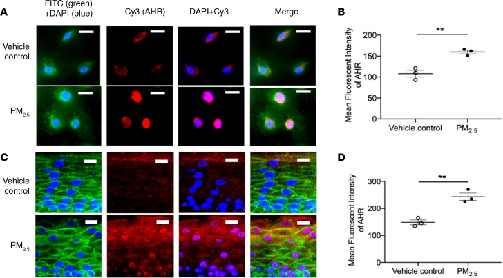
Figure 3. Effect of PM2.5 on AHR in both human primary keratinocytes and organotypic skin.
Expressions of AHR (red) in both cultured HEKs (A and B) and organotypic skin (C and D) were evaluated using immunofluorescence staining and demonstrated a reduction in FLG expression after PM2.5 exposure. Wheat germ agglutinin–conjugated FITC (green) was used to stain the cytoskeleton. Nuclei were visualized with DAPI (blue). Data are representative of 3 independent experimental repetitions. The data are shown as the mean ± SEM. n = 3 per group. Scale bar: 50 μm. **P < 0.01 by 2-tailed Student’s t test.

