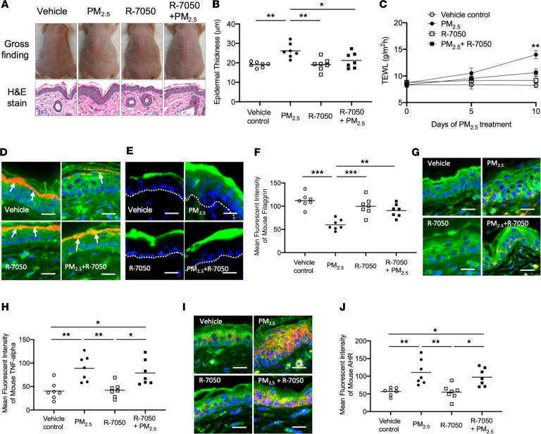
Figure 7. PM2.5 inhibits FLG and causes skin barrier dysfunction in murine skin.
Hairless mice were treated with a vehicle, PM2.5, R-7050, or a combination of PM2.5 and R-7050 on the back of each mouse twice daily for 10 days. FITC-dextran was applied to the left side of the back of each mouse for 60 minutes on day 10 and demonstrated enhanced barrier penetration of the PM2.5-treated skin. (A) Skin appearance and H&E staining (original magnification, ×100) of the skin biopsy samples in the study groups. Epidermal thickness (B) and TEWL (C) were evaluated and illustrated increased epidermal thickness and TEWL in PM2.5-exposed skin. (D) The penetration of FITC-dextran is enhanced in PM2.5-treated skin and is attenuated by TNF-α inhibitors. Protein expressions of FLG (E and F), TNF-α (G and H), and AHR (I and J) were evaluated using immunofluorescence staining. Arrows point to FLG staining (red). The dotted line represents a border between epidermis and dermis. Wheat germ agglutinin–conjugated FITC (green) was used to stain the cytoskeleton. Nuclei were visualized with DAPI (blue). Data are representative of 2 independent experiments. The data are shown as the mean ± SEM. Each point indicates individual mice, n = 7 mice per group. Scale bar: 25 μm. *P < 0.05, **P < 0.01, ***P < 0.001 by 1-way ANOVA with Tukey-Kramer post hoc test.

