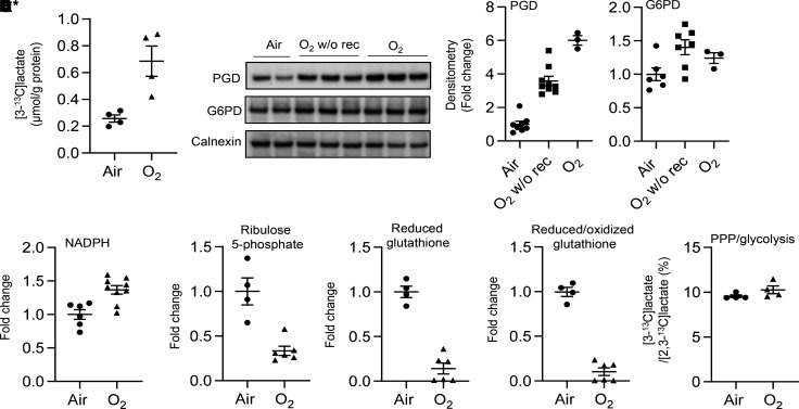
Figure 2. Hyperoxic exposure increases the PPP in cultured lung ECs.
(A–E) MFLM-91U cells (A and E) and primary LMVECs (B–D) were exposed to hyperoxia for 24 hours and then cultured in normoxia for 24 hours (refers to O2) unless specifically mentioned. (A) [3-13C]lactate was measured by the NMR when cells were incubated with [1,2-13C]glucose (20 mM) for 24 hours during air recovery phase. n = 4 per group. (B) Western blot was performed to determine levels of PGD and G6PD proteins. O2 w/o rec refers hyperoxic exposure for 24 hours without air recovery, while O2 refers hyperoxic exposure for 24 hours, followed by air recovery for 24 hours. n = 6 in air, n = 7 in hyperoxia without air recovery, and n = 3 in hyperoxia with air recovery. (C) NADPH levels were measured using a commercially available kit. n = 6 in air and n = 9 in hyperoxia. (D) Levels of ribulose 5-phosphate, reduced and reduced/oxidized glutathione were determined through metabolomics analysis. n = 4 in air and n = 6 in hyperoxia. (E) Ratio of [3-13C]lactate to [2,3-13C]lactate was calculated based on results from Figure 1D and A. n = 4 per group. Data are expressed as mean ± SEM. *P < 0.05, **P < 0.01, ***P < 0.001 versus air using 1-tailed t test (A, C, D, and E) or ANOVA followed by Tukey-Kramer test (B).

