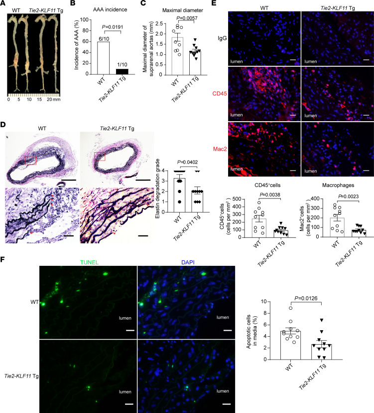
Figure 3. EC-selective overexpression of KLF11 attenuates Pcsk9/AngII-induced AAA.
The Pcsk9/AngII-induced AAA model was performed on 10-week-old male EC-selective KLF11 transgenic mice (Tie2-KLF11–Tg, n = 12) and littermate control mice (WT, n = 13). (A) Representative morphology of aortas from AngII-infused WT and Tie2-KLF11–Tg mice. (B) Incidence of AAA. (C) Maximal diameters of SAAs. (D) Representative VVG staining and quantification of elastin degradation in SAAs from WT and Tie2-KLF11–Tg mice (n = 10/group). Scale bar: 200 μm for whole aortic sections, 20 μm for magnified areas. (E) Representative immunofluorescence staining and quantification of leukocyte (CD45+) and macrophage (Mac2+) infiltration in the aortic wall of SAAs from WT and Tie2-KLF11–Tg mice (n = 10/group). Scale bar: 20 μm. (F) Representative TUNEL staining (green) and quantification of apoptotic cells in the media of SAAs from WT and Tie2-KLF11–Tg mice (n = 10/group). Scale bar: 20 μm. Data are presented as mean ± SEM. χ2 test (B), Student’s 2-tailed t test (C, E, and F), Mann-Whitney U test (D).

