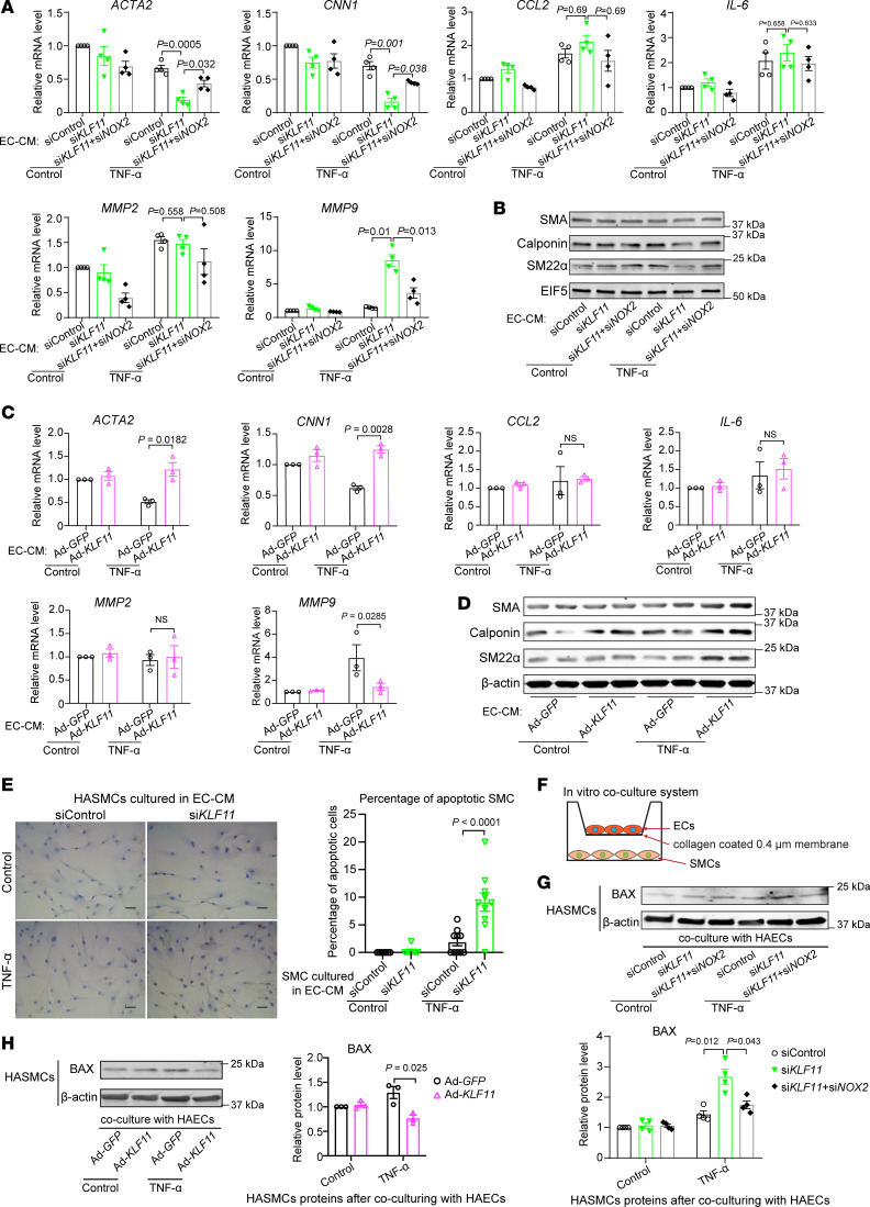
Figure 6. Coculture with KLF11-deficient ECs impairs SMC homeostasis.
(A–E) Human aortic smooth muscle cells (HASMCs) were treated for 24 hours with the conditioned media from ECs (EC-CM) that had been transfected with siControl, siKLF11, or siKLF11+siNOX2 (20 μM), or infected with Ad-GFP or Ad-KLF11 (10 MOI) and subsequently stimulated for 1 hour with TNF-α (2 ng/mL) 48 hours after siRNA transfection or adenovirus infection and cultured in fresh opti-MEM for an additional 4 hours. (A–D) qPCR (A and C) and Western blot (B and D) to examine expression of SMC-specific contractile markers (smooth muscle α-actin [SMA], calponin, and smooth muscle 22–α [SM22α]), proinflammatory cytokines (MCP-1 and IL-6), and metalloproteinases (MMP2 and MMP9). (E) HASMCs were cultured in EC-CM for 48 hours, followed by immunostaining of TUNEL. Scale bar: 20 μm. (F–H) Schematics of the in vitro coculture system using a Transwell. HAECs (upper chamber) transfected with siControl, siKLF11 (20 μM), or siKLF11+siNOX2 or infected with Ad-GFP or Ad-KLF11 (10 MOI) were cultured for 48 hours followed by TNF-α (2 ng/mL) stimulation for 1 hour separately from HASMCs, changed to fresh opti-MEM, and then cocultured with HASMCs (bottom) in fresh opti-MEM for 24 hours. The expression of BAX in HASMCs was assayed by Western blot (G and H). Data are mean ± SEM from 3 independent experiments. Two-way ANOVA followed by Holm-Sidak post hoc analysis (A, C, E, G, and H).

