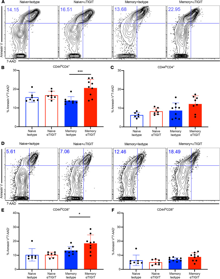
Figure 3. Apoptosis of CD44hi memory T cells is accelerated by αTIGIT Ab in memory but not previously naive septic mice.
Memory mice and age-matched naive controls received CLP, followed by injection of αTIGIT Ab or isotype control Ab at 12 and 24 hours after CLP. Mice were sacrificed and spleens were harvested at 48 hours after CLP. Splenocytes were stained with annexin V and 7-AAD for T cell apoptosis by flow cytometry. (A) Representative flow plots for annexin V+ and 7-AAD– staining gated on CD44hiCD4+ T cells. (B and C) Summary data depicting frequency of apoptotic (annexin V+ 7-AAD–) CD44hiCD4+ and CD44loCD4+ T cells in previously naive versus memory mice treated with αTIGIT Ab or isotype Ab (n = 7–9/group). (D) Representative flow plots for annexin V+ and 7-AAD– staining gated on CD44hiCD8+ T cells. (E and F) Summary data of frequency of apoptotic CD44hiCD8+ and CD44loCD8+ T cells among the 4 groups (n = 7–9/group). Groups were compared using 1-way ANOVA analysis and Tukey’s multiple comparison test. *P < 0.05, ***P < 0.001.

