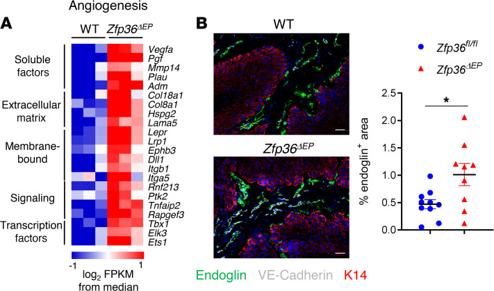
Figure 5. Increased neovascularization of Zfp36ΔEP papillomas.
(A) Heatmap of expression levels of angiogenesis-related genes significantly increased in Zfp36ΔEP tumor cells compared with their WT counterparts. (B) Sections of papillomas (3-mM diameter) from Zfp36ΔEP and Zfp36fl/fl mice after 12 weeks of treatment. Cryosections were stained with endoglin (green), VE-Cadherin (gray), Keratin14 (red), and nuclei (blue). The relative surface of endoglin-positive staining in papilloma sections was measured and graphed (mean ± SEM, n = 9–10). Statistical significance (*P < 0.05) was assessed by 2-tailed Mann-Whitney test.

