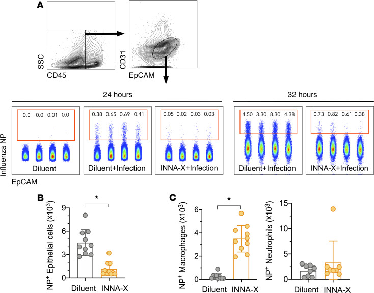
Figure 6. In vivo protection of nasal epithelial cells after viral challenge of mice treated with INNA-X.
Mice were inoculated with 1 nmol of INNA-X or diluent and 1 day later challenged with 500 PFU of Udorn IAV. Nasal turbinates were harvested 24 or 32 hours after infection and cell populations analyzed for intracellular influenza virus nucleoprotein (NP) expression. (A) Epithelial cells were distinguished by CD45–CD31–EpCAM+ expression. Center panels depict NP+-expressing cell populations from individual animals in a representative experiment. (B) Bar graphs indicate the total number of NP+ epithelial cells, (C) NP+ macrophages, or NP+ neutrophils obtained from 8–10 animals (data pooled from 2 independent experiments). All statistical analysis was performed using a Mann-Whitney t test. *P < 0.001.

