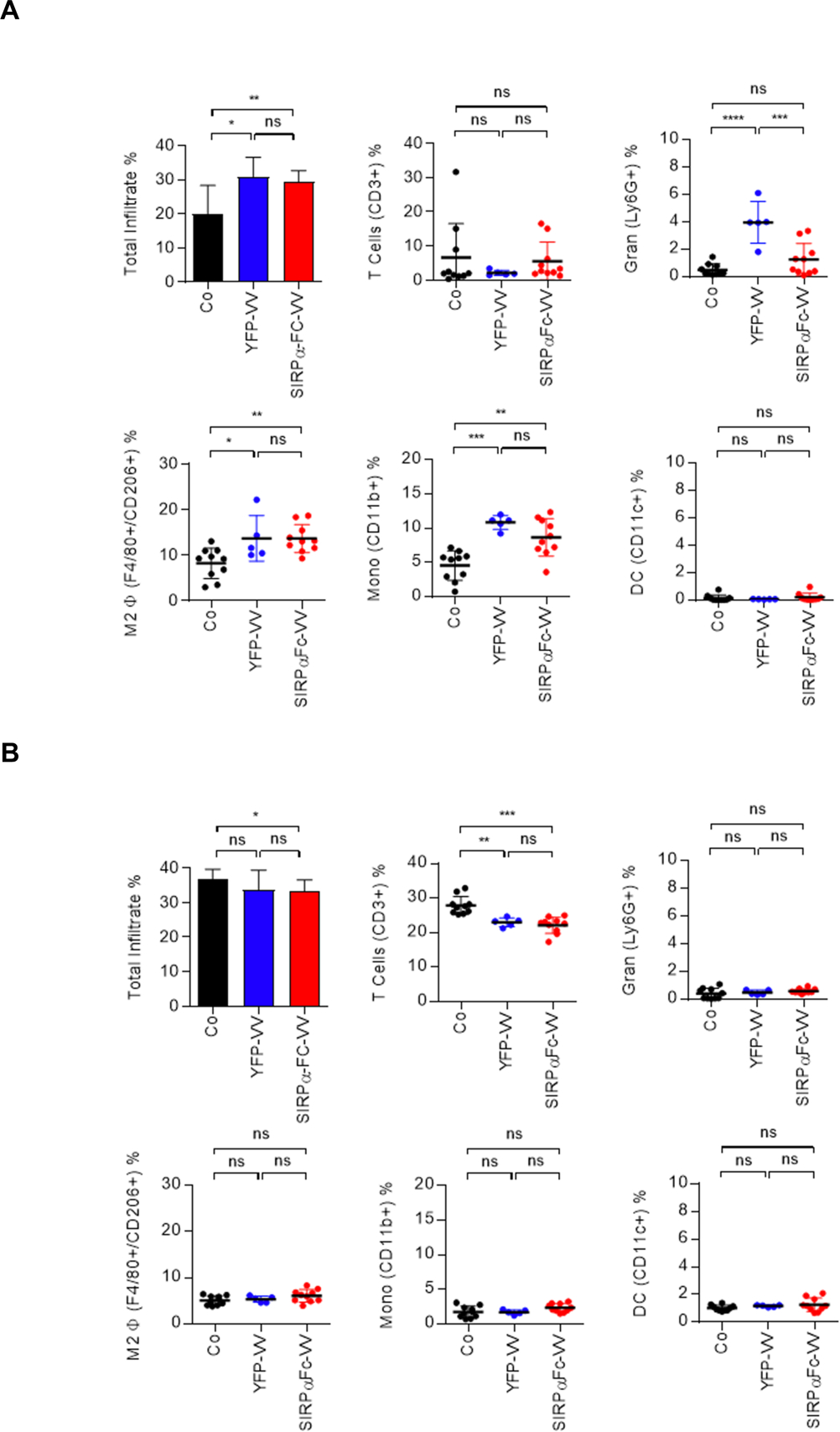
Figure 5: Changes in myeloid cell immune infiltrate in F420 tumors after VV administration.

C57BL/6 mice were injected with 1×10⁶ F420 cells s.c. into the right flank. Once the tumor was ~300mm3, 1×10⁸ PFU of SIRPα-Fc-VV (n=10) or YFP-VV (n=5) were injected i.t. PBS injected mice (n=10) served as controls (Co). After 48 hours mice were euthanized, and single cell suspensions of (A) tumors and (B) spleens were prepared. The presence of T cells (CD3+), granulocytes (Ly6G+), M2 macrophages (F4/80+, CD206+), monocytes (CD11b+), and dendritic cells (CD11c+) was determined by FACS analysis (dot: individual mice; mean percent positive +/−SD; ANOVA; *p<0.05; **p<0.01; ***p<0.001; ****p<0.0001).
