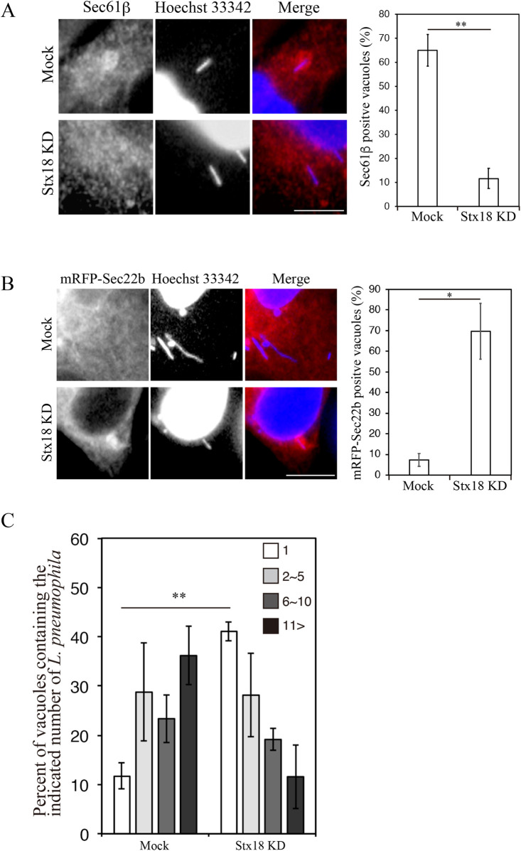
Fig 5. Stx18 is required for the LCV-ER association/fusion and intracellular Legionella growth.
(A) HeLa-FcγRII cells were transfected without (mock, top row) or with siRNA targeting Stx18 (bottom row). At 48 h after transfection, the cells were infected with L. pneumophila for 4 h, fixed, and stained with an anti-Sec61β antibody and Hoechst 33342. Bar, 5 μm. The graph shows the percentage of vacuoles positive for Sec61β. Vales are the mean ± SD (n = 3, 100 vacuoles were scored in each experiment). **P < 0.01 (Student’s t test). (B) HeLa-FcγRII cells were transfected without (mock, top row) or with siRNA targeting Stx18 (bottom row). At 24 h after transfection, the cells were additionally transfected with a plasmid for mRFP-Sec22b for 24 h, infected with L. pneumophila for 6 h, fixed, and stained with Hoechst 33342. Bar, 5 μm. The graph shows the percentage of vacuoles positive for mRFP-Sec22b. Values are the mean ± SD (n = 3, 100 vacuoles were scored in each experiment). *P < 0.05 (Student’s t test). (C) HeLa-FcγRII cells were transfected without (mock) or with siRNA targeting Stx18. At 48 h after transfection, the cells were infected with L. pneumophila for 8 h at MOI 10. Intracellular growth of L. pneumophila was scored by counting bacteria residing in a single vacuole. The graph shows that percentage of vacuoles containing 1 bacterium (white bars), 2–5 bacteria (light gray bars), 6–10 bacteria (dark gray bars), and >11 bacteria (black bars). Values are the mean ± SD (n = 3, 100 vacuoles were scored in each experiment). *P < 0.05 (Student’s t test).

