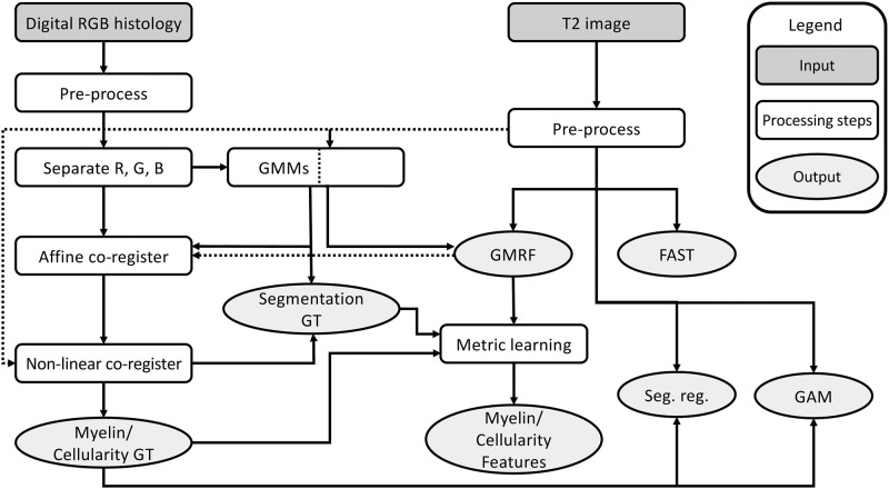
Fig 1. Image processing and analysis pipeline.
Shown are the main steps involved in this study for image preparation, MRI and histology alignment, and myelin and cellularity estimation using MRI. Note: RGB: red, green, blue; Myelin/cellularity GT: myelin and cellularity value ground truth; GMM: Gaussian mixture model; Segmentation GT: segmentation ground truth of histology; GMRF: Gaussian Markov random field; Seg. reg: segmentation regression; GAM: generalized additive model.

