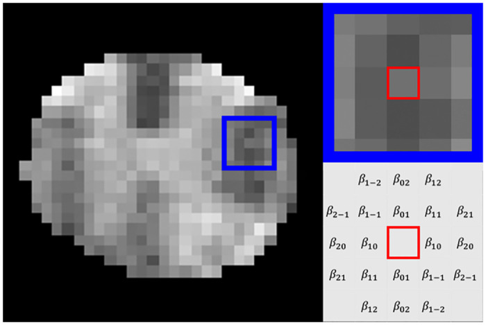
Fig 2. Markov random field model.

Shown are an example T2-weighted MR image from a mouse spinal cord (left), a focal highlight in the left lateral white matter sized 5x5 pixels (blue square, left and top right), and the Markov model parameters for an arbitrarily selected pixel within the blue (red square, bottom right). The pixel was modelled as a function of its 20 nearest neighbours, with outcomes scaled by the 10 unique, symmetric parameters.
