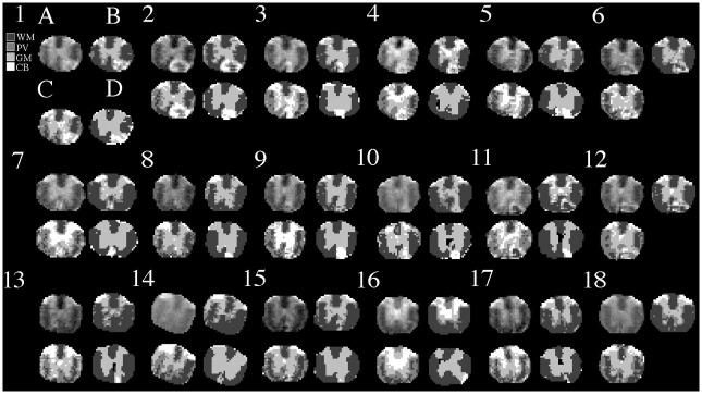
Fig 4. MRI segmentation.
Shown are the original T2 MRI images (A) from each animal (numbers), the corresponding segmentation results of MRI using our GMRF (B) and the open-source software FSL FAST (C), and the associated histology segmentation using our GMM method for comparison (D). Row indicates time cohort: day 7 (row 1), day 14 (row 2) or day 28 (row 3).

