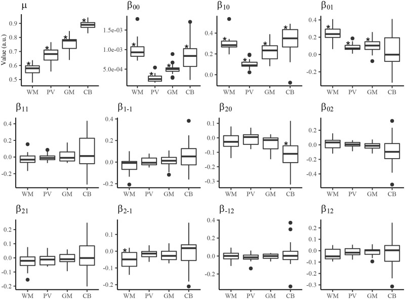
Fig 5. GMRF MRI segmentation parameters.
Shown are boxplots of the GMRF segmentation parameters across all subjects. Parameters showing no overlap of the inter-quartile ranges between tissues (WM, PV, GM, CB) suggest tissue-dependent and approximately independent of the test subject. The symbol, ⋆, indicates parameters significantly different from zero based on Bonferroni-corrected Wilcox test. Note: WM: white matter; PV: partial volume; GM: gray matter; CB: cell body.

