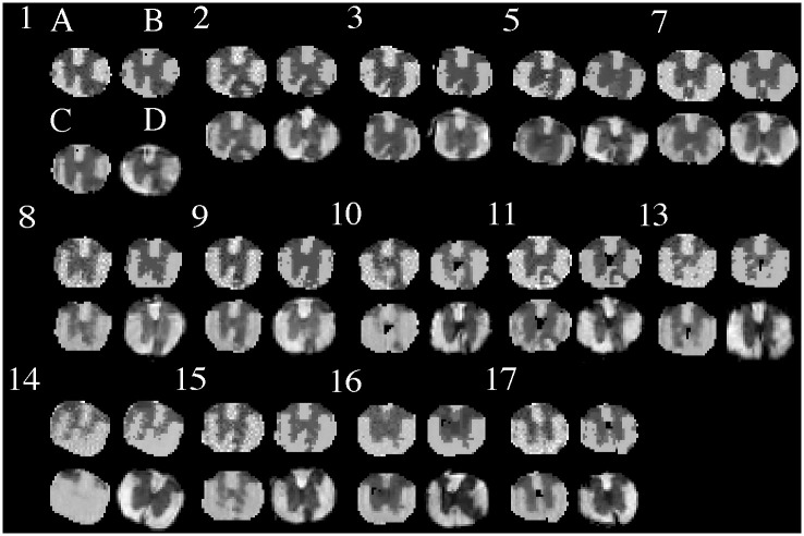
Fig 6. Myelin predictions.
Shown are the myelin feature images learned from the MRI of the 14 mice that have histological correspondence using: our GMRF (A), segmentation regression associated with our GMRF (B), Markov GAM using a freely available software (C), and the histological standard (D). Note: there is no clear myelin/WM structure detected using Markov GAM in mice 10 and 14, and the lesion cores appear to be overestimated in mice 2 and 10 in the MRI methods.

