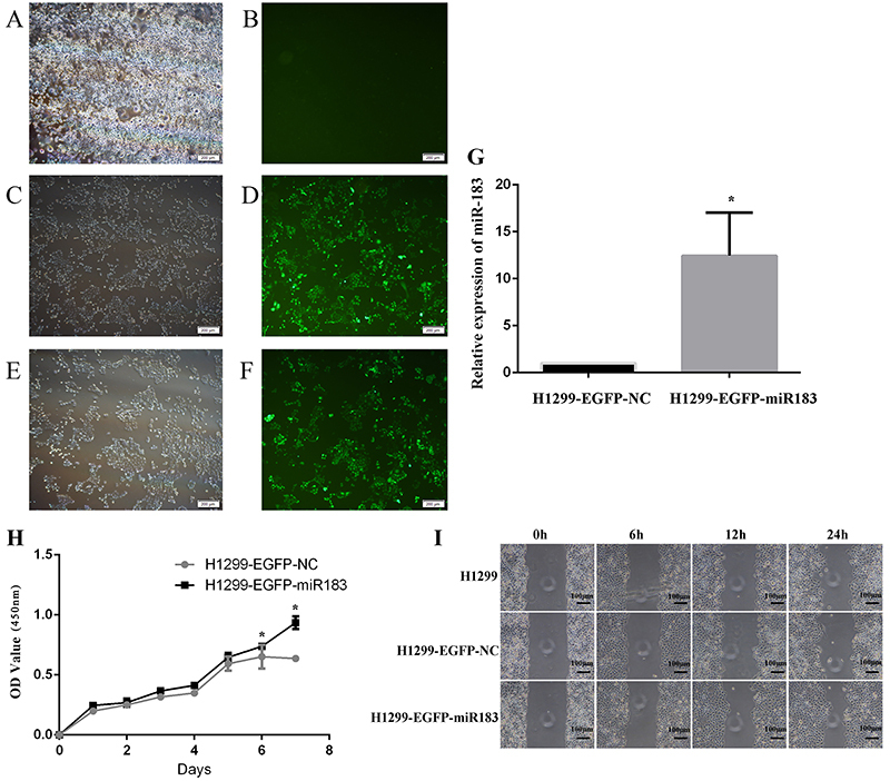
Figure 6. A-F, The expression of EGFP under a fluorescence microscope after H1299 cells were infected with lentivirus (A and B, H1299 blank control group; C and D, H1299 negative control group; E and F, H1299 miR-183-overexpression group) (scale bar: 200 μm). G, The expression of miR-183 was detected by qPCR after transfection with lentivirus in H1299 cells. H, The effect on cell proliferation after H1299 cells were infected with virus (H1299-EGFP-NC: H1299 negative control group; H1299-EGFP-miR183: H1299 miR-183-upregulated expression group). Data are reported as means±SD (n=3). *P<0.05 (t-test). I, Scratch healing test after H1299 cells were infected with virus.

