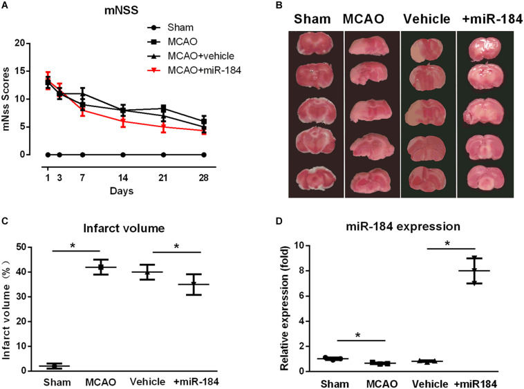
FIGURE 1.
The effect of expression levels of miR-184 in brain on the injured brain following ischemia stroke. Rats were subjected to middle cerebral artery occlusion (MCAO) operation to induce ischemic stroke model with or without intracerebroventricular injection of miR-184 adenovirus (Ad-miR-184). After 24 h following MCAO, a cohort of rats was sacrificed, and brain tissue was taken for TTC staining and qRT-PCR to evaluate the infarct volume and miR-184 expression in brain. Other rats were subjected to analysis of neurological function deficit 1, 3, 7, 14, 21, and 28 day after MCAO operation using mNSS system. (A) mNSS scores in various groups (n = 8–9). (B) TTC-stained brain slice images 24 following ischemia-reperfusion (n = 6 in each group). (C) Quantification analysis of the infarct volume. (D) Relative expression of miR-184 in the brain (n = 6 in each group). Values are mean ± SD. *p < 0.05 vs. the sham or vehicle.

