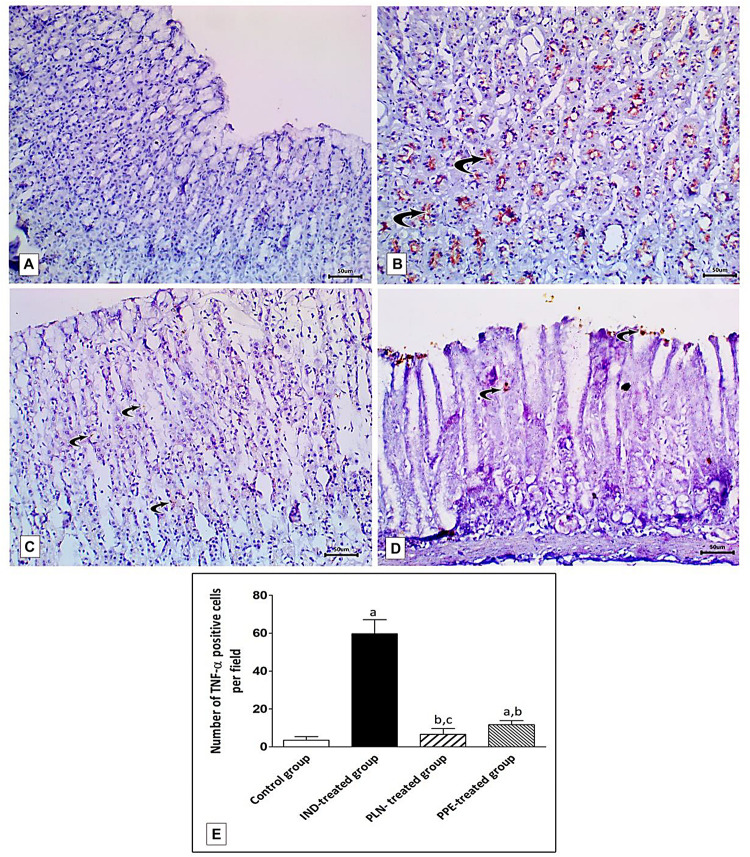
FIGURE 12.
Sections from the fundic mucosa demonstrating immunohistochemical reactivity to TNF-α in the different studied groups. (A) the control group shows weak TNF-α immunoreaction in the gastric mucosa. (B) IND-treated group shows highly positive TNF-α immunoreaction especially in the basal glands (curved arrow). (C) PLN-treated group shows weak positive TNF-α immunoreaction (curved arrow). (D) PPE-treated group showing mild positive TNF-α immunoreaction especially in surface epithelium (Immunostaining for TNF-α × 200, scale bar = 50 μm). (E) quantitative analysis of the mean count of TNF-α immunopositive cells in the fundic mucosa of the studied groups. Data are presented as mean ± SD. Kruskal-Wallis H test followed by the Mann-Whitney U tests were used for analyzing the results. asignificant as compared to the control group (p ≤ 0.05). bsignificant as compared to IND-treated group (p ≤ 0.05), csignificant as compared to PPE-treated group (p ≤ 0.05).

