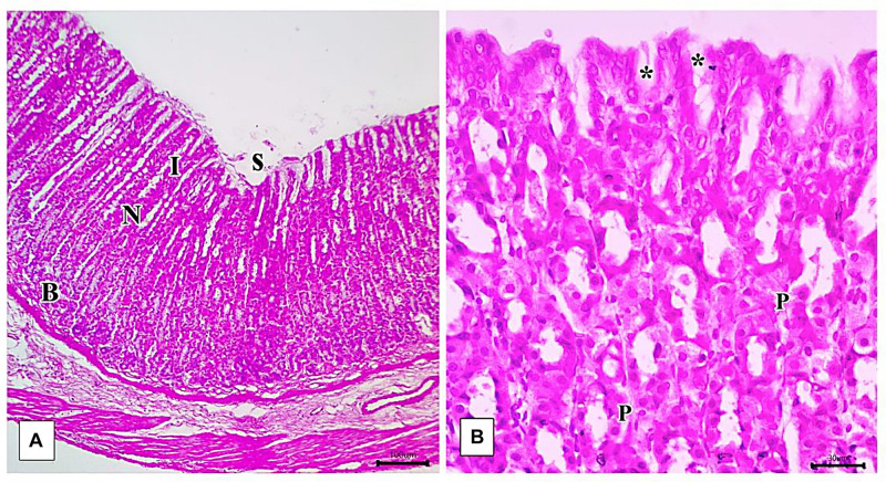
FIGURE 8.
Photomicrographs of the fundic mucosa of the control group illustrating (A) normal histologic structure of surface lining epithelium (s), longitudinally arranged healthy-looking glands consisting of the isthmus (I), neck (N), body, and base (B) (H&E × 100, scale bar = 100 μm). (B) the upper part of the fundic gland surface epithelial columnar mucous secreting cells (s) short gastric pits (*). Notice that parietal cells are distinguished by their rounded nuclei and acidophilic cytoplasm (P) (H&E × 400, scale bar = 30 μm).

