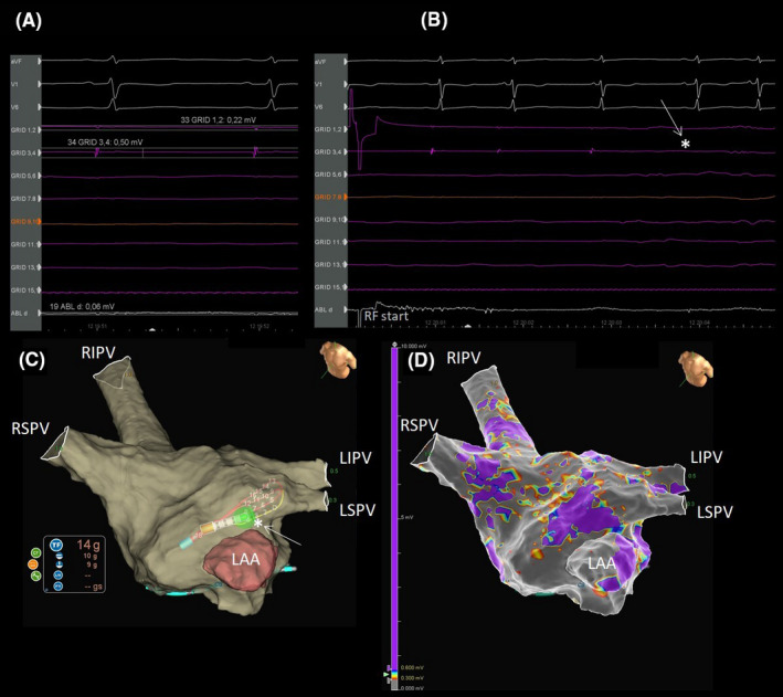
FIGURE 2.

Direct comparison of bipolar versus omnipolar recordings. A, Pair 3, 4 of the HD Grid records a 0.5 mV signal, while the signal recorded by pair 1, 2 is of 0.22 mV. The distal dipole of the ablation catheter records a signal of 0.06 mV. B, During PVI complete disappearance of the HD Grid signal at the fourth beat after the beginning of RF (white arrow and star). C, The 3d anatomical map. Note the contact force of the ablation catheter equal to 14 grams during PVI (white arrow and star). D, The related electro‐anatomical map.
