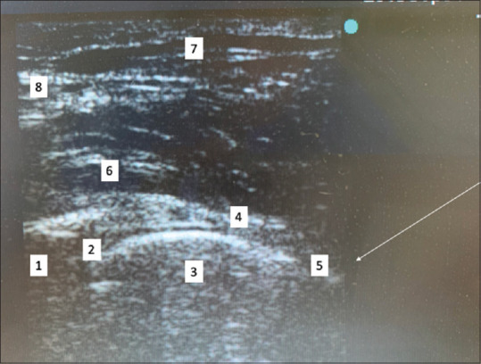
Figure 3.

Ultrasound image showing hip joint injection. Longitudinal view high-frequency ultrasound linear probe. 1 acetabulum, 2 hyperechoic labrum, 3 femoral head, 4 iliofemoral ligament, 5 anterior recess, 6 iliopsoas, 7, sartorius. The white line represents the trajectory of the needle for placement in hip joint
