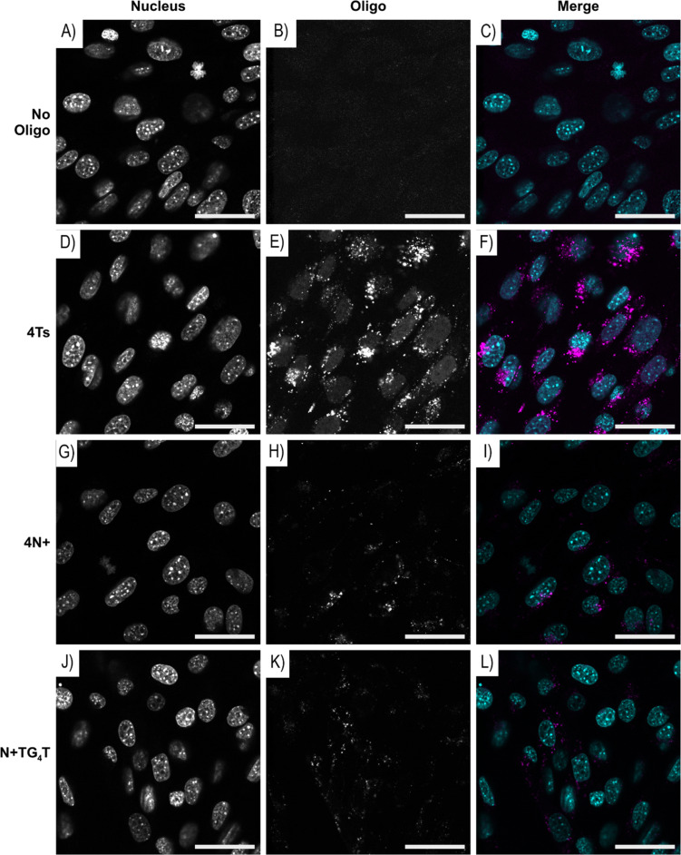
Figure 3.
Representative images of mouse NIH 3T3 fibroblasts incubated with either (A–C) no oligo or 20 µM of (D–F) 4Ts, (G–I) 4N+, and (J–L) N+TG4T FAM labelled ONs. Asynchronously growing NIH3T3 cells were incubated for 12 hours with 20 µM of the stated FAM-labelled ONs or without ON, then fixed with 4% paraformaldehyde before staining with Hoechst 3342 to identify nuclear DNA. The images were collected with a Leica SP5 DM6000B scanning confocal microscope. Individual panels, nucleus/Hoechst 3342 and oligo/FAM are shown for each section, along with merge where pseudo-coloured panels are overlaid, nucleus (blue) and oligo (magenta). Scale bar: 40 μm.

