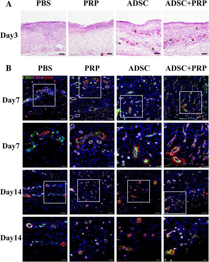
Fig. 4.
Evaluation of blood vessel formation. a Early angiogenesis in the wound beds was visualized on day 3 postwounding by hematoxylin and eosin staining. Staining showed an increased number of blood vessels forming in the wound in the ADSC+PRP group compared with the other groups. The black arrows indicate blood vessels. Scale bars = 50 μm. b Representative images of tissue sections from diabetic rats immunostained for CD31 (green), CD34 (pink), and α-SMA (red) and quantification of c the capillary density and the proportion of CD31+, CD34+, and α-SMA+ cells 7 and 14 days postwounding (× 200, scale bars = 50 μm; × 400, scale bars = 20 μm; *p < 0.05; n = 3 wounds per group)

