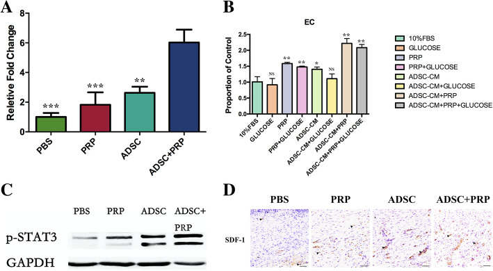
Fig. 5.
Effects of ADSC+PRP in promoting angiogenesis and vasculogenesis. a ELISA of VEGF in skin tissue lysates postwounding showed higher levels of VEGF protein production in the wounds of the ADSC+PRP group than those of the other groups. All experiments were performed in triplicate and were repeated three times to confirm the findings. Values were expressed as mean ± SEM. Statistical analysis was evaluated using one-way ANOVA and Tukey post hoc test (significance was set to *p < 0.05, **p < 0.01, and ***p < 0.001). b ADSC+PRP significantly enhanced the proliferation of endothelial cells compared with the ADSC-CM, PRP, or control group; the similar phenomenon was also presented in high-glucose medium. All experiments were performed in triplicate and were repeated three times to confirm the findings. Values were expressed as mean ± SEM. One-way ANOVA and Tukey’s post hoc test showed statistically significant differences overall between the eight groups (significance was set to *p < 0.05, **p < 0.01, and ***p < 0.001). c Protein expression levels of p-STAT3 on wound tissues were detected by Western blot analysis. d Representative images of tissue sections from diabetic rats immunostained for SDF-1 14 days postwounding (× 400, scale bars = 20 μm; *p < 0.05; n = 3 wounds per group)

