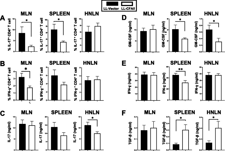
Fig. 7.
CD4+ T cell SjS recipients from LL-CFA/I-treated donors showed reduced proinflammatory cytokine production and increased TGF-β production. Recipient lymphocytes isolated from the MLNs, spleens, and HNLNs were restimulated with anti-CD3 plus anti-CD28 mAbs. a, b After 2 days, lymphocytes were harvested for flow cytometry analysis to measure percent a IL-17+ and b IFN-γ+ CD4+ T cells. Collected cell culture supernatants from 4-day restimulated cultures were analyzed for the presence of c IL-17, d GM-CSF, e IFN-γ, and f TGF-β. *P < 0.05, **P < 0.01, differences in recipients given donor CD4+ T cells from LL vector- or LL-CFA/I-treated SjS mice are shown

