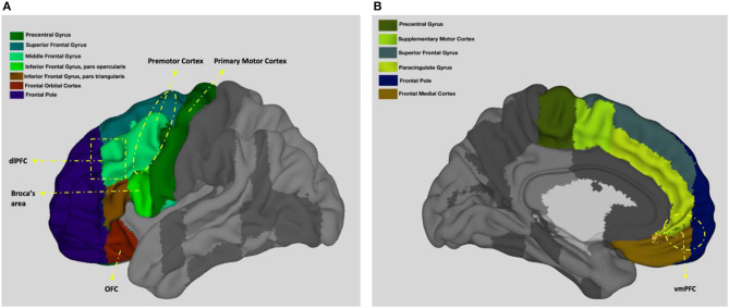
Figure 2.
Neuroanatomy of the frontal lobes. (A) presents left view and (B) presents right view. Adapted from the Harvard-Oxford atlas developed at the Center for Morphometric Analysis (CMA), and distributed with the FMRIB Software Library (FSL) (Bakker et al., 2015), 3D Surface View (Majka et al., 2012).

