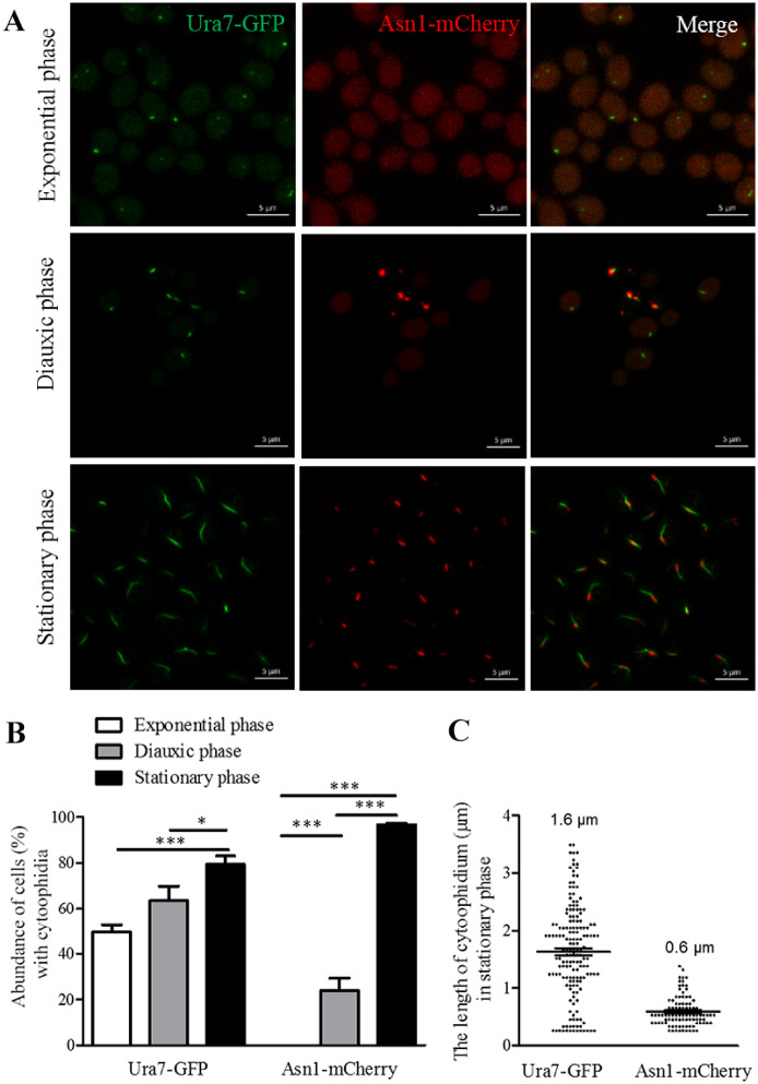
Figure 1.
Ura7 and Asn1 cytoophidia in S. cerevisiae. Ura7-GFP Asn1-mCherry cells were grown in rich medium and cells were collected after 6, 24, and 168 h culture. The cells were subsequently fixed by 4% PFA at room temperature for 10 min. Ura7-GFP and Asn1-mCherry protein were observed by fluorescent microscopy. The abundance of cytoplasmic cytoophidia were calculated and the average length of cytoplasmic cytoophidia were plotted. (A) Representative confocal image of Ura7-GFP Asn1-mCherry cells. Ura7 cytoophidia were observed in all three growth phases. Scale bar 5 μm. (B) Quantification of cells with visible cytoophidia was plotted and expressed as percentage of cells containing cytoophidia in three growth phases. *P < 0.05 and ***P < 0.001 (C) The average length of Ura7 and Asn1 cytoophidia was measured and plotted in the stationary phase.

