Fig. 6.

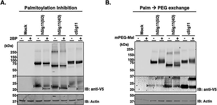
Palmitoylation of Siglec-11. (A) V5-tagged Siglec-11 stably transfected HEK293 cells were cultured with or without protein acylation inhibitor 2BP and the cell lysates were analyzed by western blotting with anti-V5 tag antibody. β-actin was used as loading control. Arrows indicate molecular weight shift as the result of 2BP. (B) Protein S-fatty acylation was tagged by 5 kDa-PEG with cell lysates of V5-tagged Siglec-11 stably transfected HEK293 and analyzed by western blotting with anti-V5 tag antibody. Expression level of full length Siglec-11 (higher molecular weight membrane) and proteolytical fragment (lower molecular weight membrane) were different, thus different exposures were needed. Arrows indicate molecular weight shift in the presence of PEG tag.
