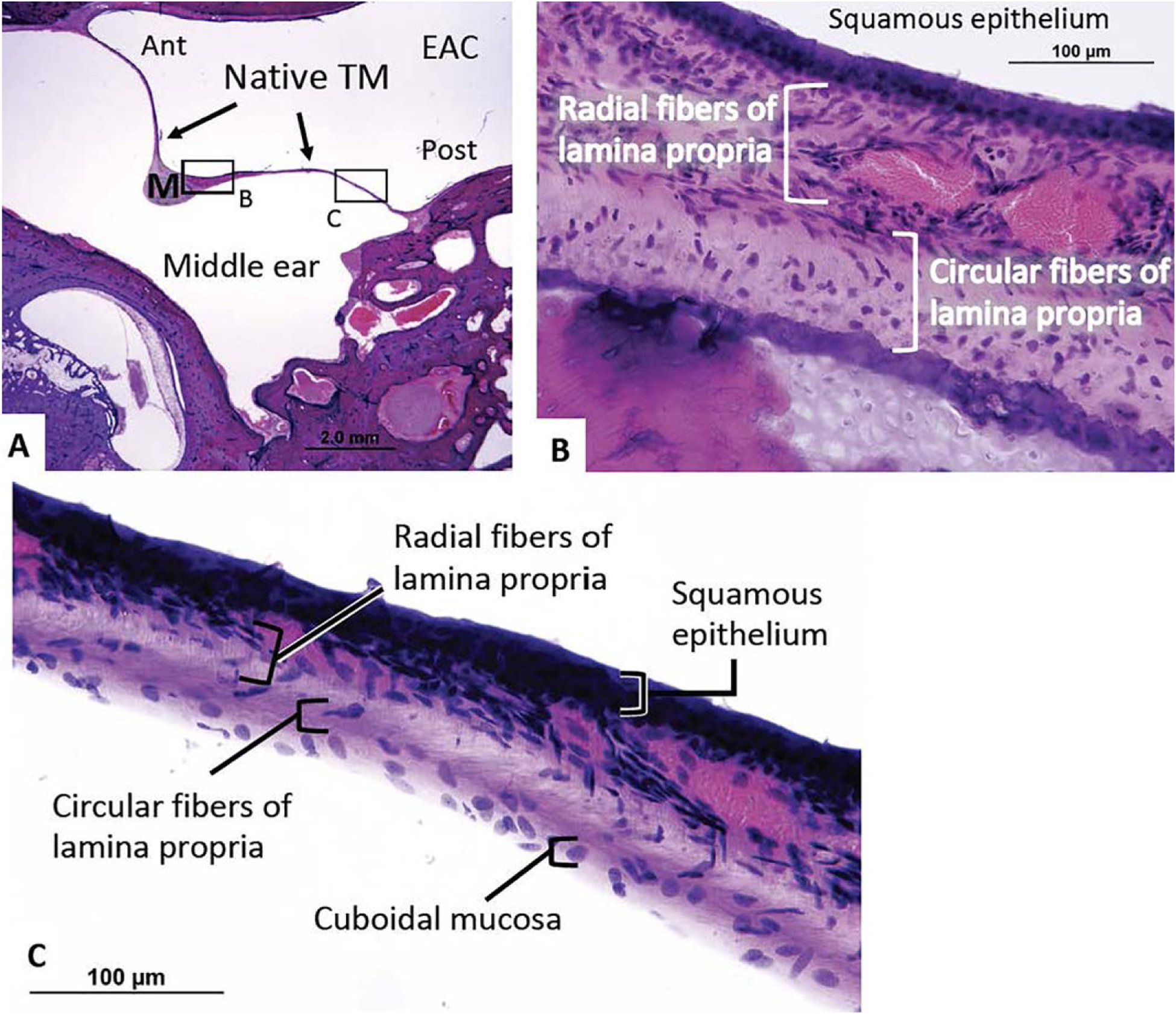
Fig. 6.

(A) A representative healthy left TM from a 77-year-old patient without history of middle ear disease or surgery. (B) There is a clear pattern of outer radial fibers and inner circular fibers within the lamina propria. (C) This thin radial and circular fiber pattern is easily seen throughout the TM.
Ant = anteriorly; EAC = external auditory canal; M = malleus; Post = posteriorly; TM = tympanic membrane.
