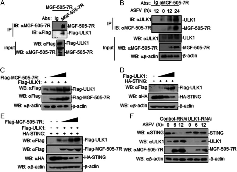
FIGURE 7.
ASFV MGF-505-7R degrades STING by upregulating ULK1 expression. (A) MGF-505-7R interacted with ULK1 in the overexpression system. 293T cells (2 × 10⁶) were transfected with the indicated plasmids (5 μg each). Coimmunoprecipitation and immunoblotting analyses were performed with the indicated Abs. The expression of transfected proteins was analyzed by immunoblotting with anti–MGF-505-7R or anti-Flag Abs. (B) Endogenous associations between MGF-505-7R and ULK1. PAM cells were uninfected or infected with ASFV (MOI: 0.01) for the indicated times before coimmunoprecipitation and immunoblot analysis. (C) MGF-505-7R increased the expression of ULK1 protein. 293T cells (2 × 10⁵) were transfected with HA-ULK1 (1 μg) and Flag-MGF-505-7R (0, 0.25, 0.5, and 1.0 μg) for 24 h. Cell lysates were analyzed by immunoblotting with the indicated Abs. (D) ULK1 inhibits the expression of STING protein. The experiments were similarly performed as in (C). (E) ULK1 increases MGF-505-7R degradation of STING. 293T cells (2 × 10⁵) were transfected with the indicated plasmids for 24 h. Cell lysates were analyzed by immunoblotting with the indicated Abs. (F) Effects of ULK1 knockdown on STING and MGF-505-7R. PAM cells were transfected with control–RNAi or ULK1–RNAi (2 μg). Twenty-four hours after transfection, cells were left uninfected or infected with ASFV (MOI: 0.01) for the indicated times before immunoblotting was performed with the indicated Abs. All the experiments were repeated three times with similar results.

