Fig. 3.
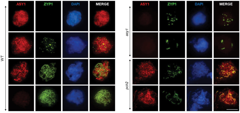
Synapsis is defective in asy1 and pch2. Immunolocalization of ASY1 (red) and ZYP1 (green) in the WT, asy1, and pch2. In asy1, ASY1 is not detected and ZYP1 forms foci in zygotene–pachytene cells and aggregates in late pachytene–diplotene cells. In pch2, different from the WT, ASY1 does not get depleted from the axes and appears highly abundant in limited synapsed regions co-localizing with ZYP1. DNA is counterstained with DAPI and shown in blue. Scale bar=10µm.

