
Fig. 1 – Site of hemolymph sampling and workflow of the library preparation and the analysis.
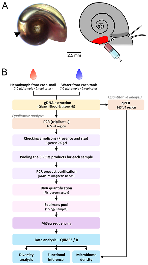
A. Snail hemolymph was sampled directly by heart puncture using a syringe. Left panel shows the position of the heart (arrow) on an albino snail. Right panel shows a schematic representation of the puncture. B. Work flow of the sample preparation and data processing.