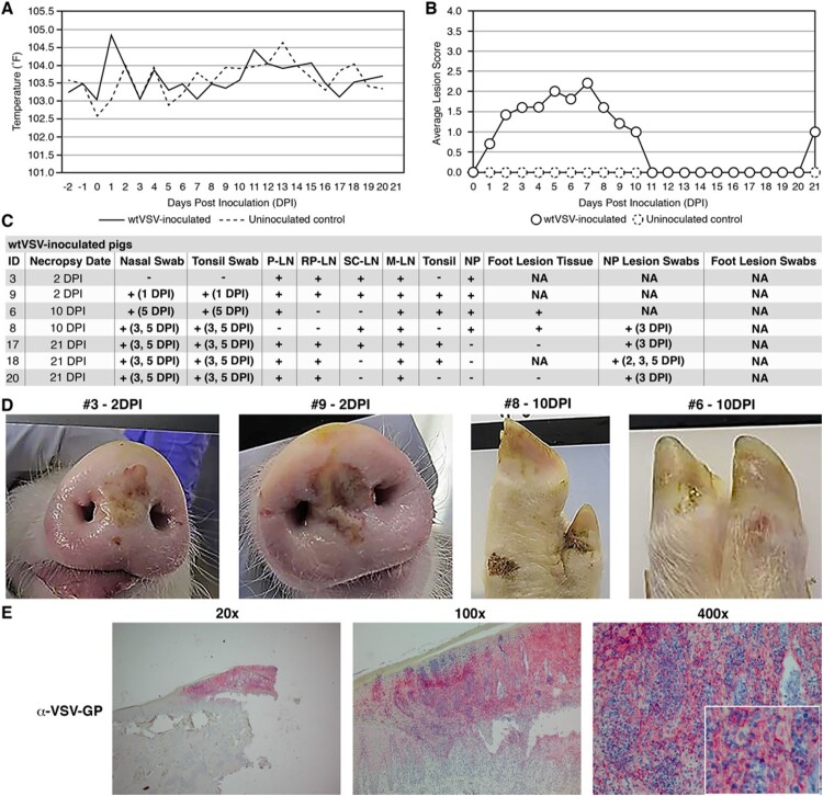
Figure 1.
Clinical analysis of wtVSV-inoculated pigs. (A) Average daily temperatures of wtVSV-inoculated pigs compared to uninoculated pigs. (B) Average lesion scores of wtVSV-inoculated pigs compared to uninoculated environmental controls on each day of the study. (C) Summary of RT-qPCR analysis performed to detect viral RNA in clinical samples collected throughout the study, indicating the presence (+) or absence (−) of viral RNA. P-LN – parotid lymph node; RP-LN – retropharyngeal lymph node; SC-LN – superficial cervical lymph node; M-LN – mandibular lymph node; NP – nasal planum; NA – Not Applicable because not present or collected; DPI - days post inoculation in which positive samples were detected. (D) Representative pictures showing vesicular lesions in pigs. (E) Immunohistochemistry analysis performed on nasal planum lesion tissue collected on 2 DPI necropsy from wtVSV-infected pig #3 showing positive (red) immunostaining using anti-VSV-G rabbit polyclonal antibody localized to the stratum spinosum and granulosum of the epidermis.

