Figure 3.
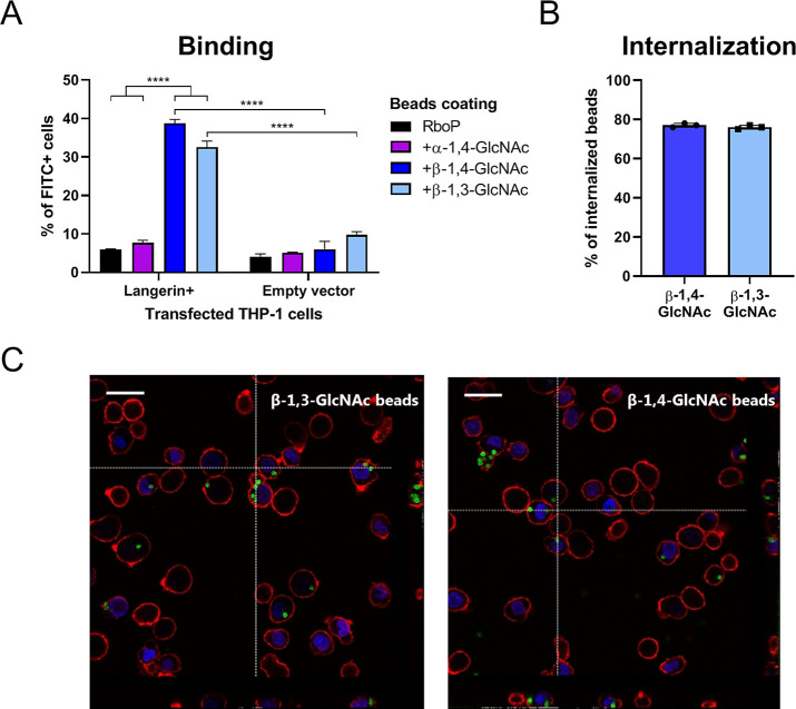
Binding and internalization of β-GlcNAc-WTA-coated beads by langerin-expressing THP-1 cells. (A) Binding of FITC-labeled beads, coated with unglycosylated or in vitro glycosylated RboP hexamers, to THP-1 cells transfected with human langerin or empty vector at a bead-to-cell ratio of 1. Adherence is represented by percent of FITC+ cells. (B) Proportion of adherent β-GlcNAc WTA beads that is internalized by Langerin + THP-1 cells. (C) Confocal microscopy images (40×) of β-GlcNAc WTA beads (FITC-labeled: green) bound to and internalized by Langerin+THP-1 cells (WGA-Alexa 647: red, DAPI: blue). Vertical lines correspond to cross section of z-stack on the right, horizontal lines to cross section below, scale bars correspond to 25 μm. For panels A and B, graphs represent mean + SEM of biological triplicates, ****p < 0.001.

