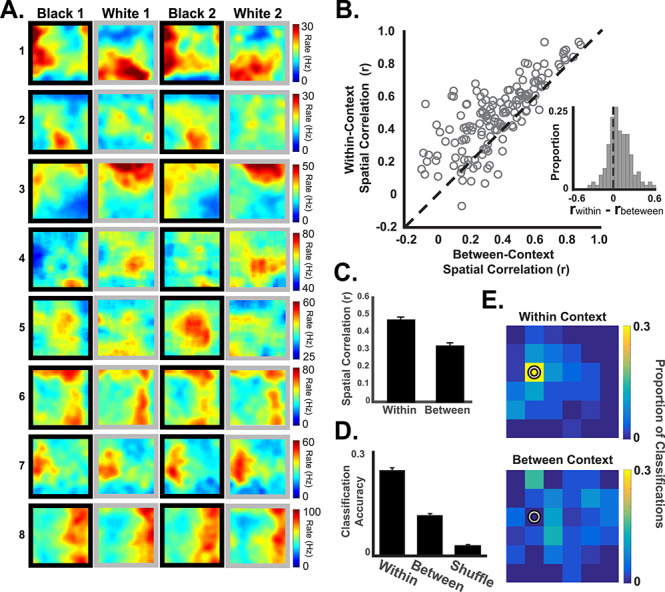
Figure 1.

Retrosplenial cortical neurons exhibit context-specific spatial coding. (A) Spatial firing rate maps from illustrative examples of spatial firing patterns in the black and white contexts. Some neurons (e.g., neurons 1–3) exhibited spatially localized firing that was context dependent, whereas others (4–5) exhibited less spatial specificity (note that the color scale starts above 0). Some neurons (e.g., 6–8) exhibited spatial firing patterns that were not context dependent. Plots are shown in Black-White-Black-White order for illustration. The actual order of black and white trials was randomized. (B) Pixel-by-pixel correlations (open circles) of firing rate maps for trials occurring in the same context (y-axis) and different contexts (x-axis). Most neurons exhibited more similar spatial firing for visits to the same context than for different contexts (circles above the unity line and inset). (C) Average within-context correlations were significantly higher than between-context correlations. (D) Decoding of the rat’s current position from population activity patterns was more accurate when the classifier was trained on data from the same context (within) than when trained on data from the different context (between) and compared to shuffled data. (E) Heat maps illustrating an example of successful decoding of the current position (pixel with a circle) using same-context data (top) as compared to different-context data. One example location is shown here. See Supplementary Figure S4 for all other locations.
