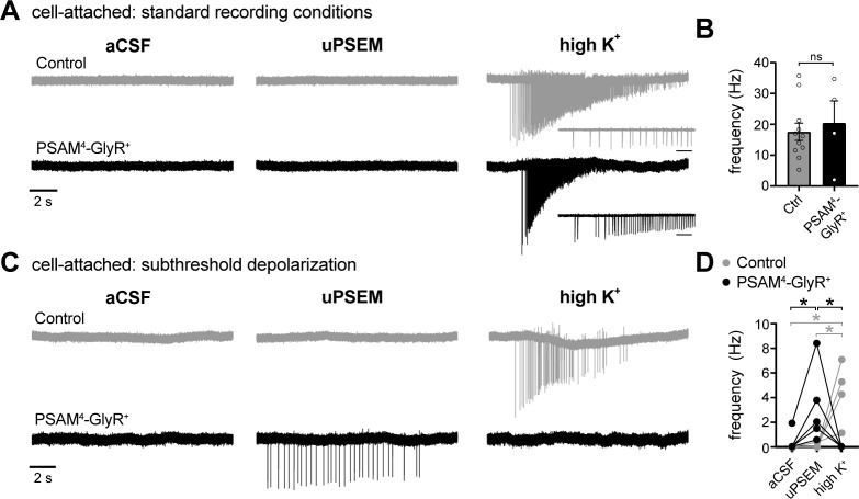
Figure 4. In cell-attached configuration, activation of PSAM4-GlyR triggers firing of D1-MSNs when the membrane is depolarized to subthreshold potentials.
(A) Representative traces of cell-attached recordings from a control (gray) or PSAM4-GlyR+ neuron in standard aCSF (4.5 mM [K+]). Action currents were evoked following uPSEM792 by applying a high external concentration of potassium (high K+, ~40 mM). The insets below the high K+ traces represent an expanded timescale at the beginning of firing; Inset scale bar = 200 ms. (B) In PSAM4-GlyR+ neurons, activation of PSAM4-GlyR with uPSEM792 had no effect on the frequency of action currents in response to high K+. (C) Representative traces of cell-attached recordings from a control (gray) or PSAM4-GlyR+ neuron in aCSF containing 10–13 mM [K+]. In PSAM4-GlyR+ neurons, activation of PSAM4-GlyR with uPSEM792 produced firing. There was no further firing with application of high K+. uPSEM792 had no effect on firing in control neurons. (D) Plot of the frequency of action currents in control and PSAM4-GlyR+ neurons in aCSF, uPSEM792 and high K+ (mixed effect two-way ANOVA with Sidak’s multiple comparisons test). Line and error bars represent means ± SEM. * indicates statistical significance, ns denotes not significant.

