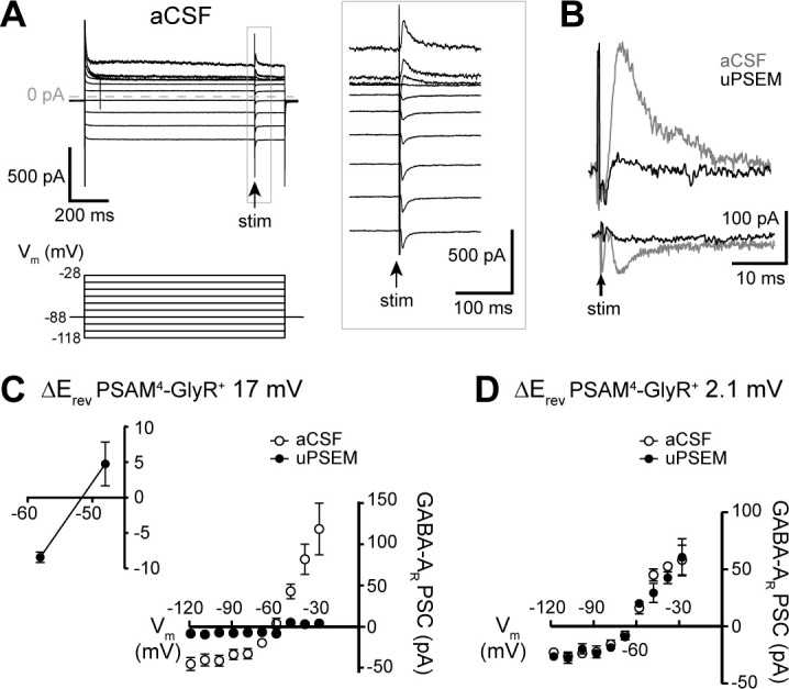
Figure 6. PSAM4-GlyR activation in D1-MSNs reduces GABAergic synaptic inhibition.

(A) Representative traces of GABA-A receptor-mediated synaptic currents in a PSAM4-GlyR+ neuron in aCSF, evoked during voltage steps from Vhold −88 mV (voltage steps: 1 s, −118 to −28 mV, 10 mV; electrical stimulation at 850 ms into voltage steps). Panel on the right shows the traces in expanded timescale corresponding to the gray rectangle on the left. (B) Representative traces of GABA-A receptor-mediated currents evoked in the same neuron from Vhold −28 mV (outward) and −118 mV (inward) in aCSF (gray) and uPSEM792 (black). (C) Plot of the current–voltage relationship of the GABA-A receptor synaptic currents in aCSF and uPSEM792 in neurons where the reversal potential of PSAM4-GlyR current shifted by >5 mV (n = 5, average shift of 17.0 mV). The reversal potential of GABA-A receptor-mediated current was −60.9 mV in aCSF and −51.6 mV in uPSEM792. Inset (top left) shows values of GABA-A receptor synaptic currents in uPSEM792 near the reversal potential in an expanded scale. (D) Plot of the current–voltage relationship of the GABA-A receptor-mediated synaptic current in aCSF and uPSEM792 in neurons where the reversal potential of PSAM4-GlyR current did not shift (n = 3, average shift of 2.1 mV). The reversal potential of GABA-A receptor-mediated synaptic currents was similar in aCSF (−64.7 mV) and uPSEM792 (−64.9 mV). Line and error bars represent means ± SEM, * indicates statistical significance.
