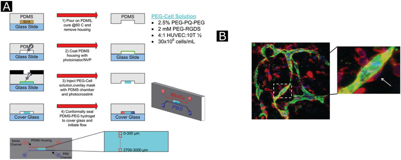
FIG. 10.
Schematics of microfabrication design (a), in step 1, to fabricate an external perfusion housing, PDMS is replica molded. In step 2, the interior of the housing is coated with a photoinitiator. In step 3, the housing is injected with photopolymerizable PEG precursors, and to fabricate hydrogel microchannels within the external PDMS housing, mask-directed photolithography is used. In step 4, the PDMS–PEG multilayer device is conformally sealed to coverglass and perfused with media and buffer. The last schematic shows the spatial relationship of the perfused media (red) and buffer (blue) microchannels to PEG hydrogel (cyan) regions imaged for analysis. Immunohistochemistry (IHC) of microvascular network formation at 96 h (B), where HUVECs (green), 10T1/2 cells (red), and nuclei (blue) Reprinted with permission from Cuchiara et al., Adv. Funct. Mater. 22, 4511 (2012). Copyright 2012 John Wiley & Sons, Inc.

