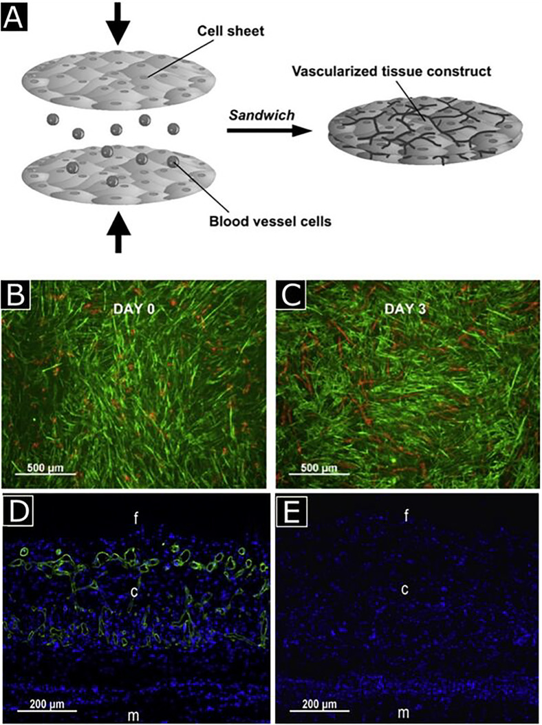
FIG. 4.
Schematic representation for vascularized tissue formation by sandwich method (a) human umbilical vein endothelial cells (HUVECs) sandwiched between myoblast sheets with the help of a gelatin-coated plunger, cultured up to 3 days, and stained with UEA-I (red) and anti-desmin antibody (green) for HUVECs and myoblasts, respectively (b) and (c). Observation of neovascularization with anti-human CD31 antibody (green) staining in five-layered myoblast sheet constructs with (d) and without HUVECs (e). Notations f, c, and m represent the fibrin gel, cell sheet construct, and muscle tissue, respectively. Reprinted with permission from Sasagawa et al., Biomaterials 31, 1646 (2010). Copyright 2010 Elsevier.

