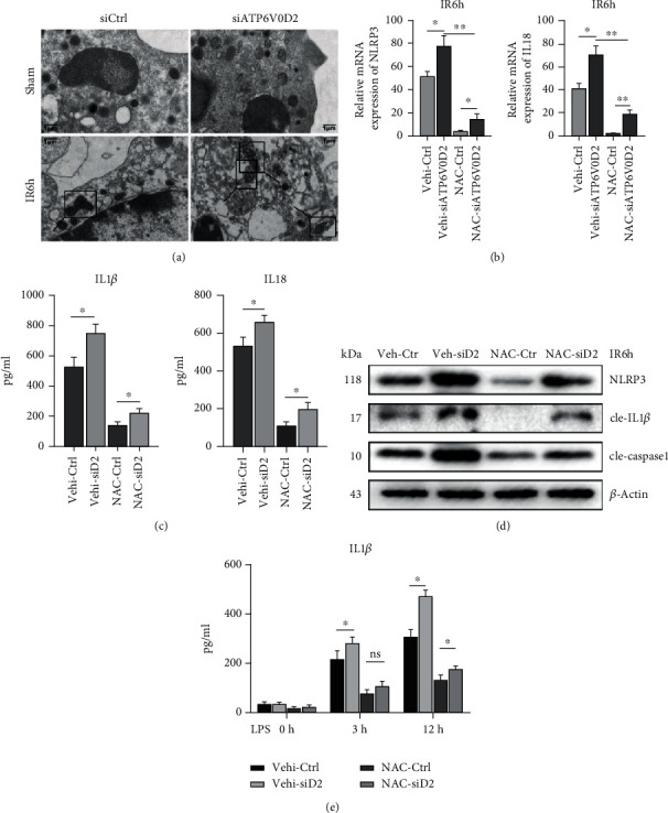
Figure 4.

Knockdown of ATP6V0D2 aggravates ROS-related mitochondrial damage after IR. (a) Mitochondria in liver macrophages were detected by transmission electron microscopy at IR6h; the areas marked by box were mitochondria (1000x magnification; scale bars, 1 μm; representative of three experiments). (b) NAC was pretreated (150 mg/kg, ip) 30 min before IR surgery, then detected the expression of NLRP3 and IL18 in macrophages harvested from siATP6V0D2 and siCtrl IR6h groups by quantitative real-time-PCR (n = 4‐6 mice/group). ∗∗P < 0.01. (c) After pretreating NAC or vehicle, isolated macrophages from siATP6V0D2 and siCtrl groups were cultured for another 6 h in vitro. IL-1β and IL-18 levels were measured in the culture supernatant by ELISA (n = 4‐6 mice/group). (d) On the basis of pretreating NAC or vehicle, the levels represented for activation of NLRP3-related proteins were detected by western blot in siATP6V0D2 and siCtrl groups at IR6h (representative of three experiments). (e) BMDMs were transfected with siATP6V0D2 or siCtrl for 24 h and then stimulated with LPS for 0, 3, and 6 h in vitro. NAC (5 mM) or ctrl was treated 1 h before LPS stimulation. IL-1β in the culture supernatant was measured by ELISA (n = 4‐6 mice/group).
