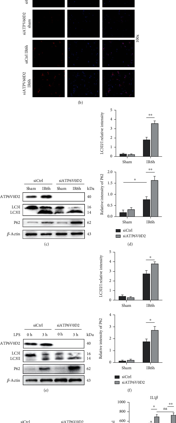
Figure 5.

siATP6V0D2 induces NLRP3 activation by impairing IR autophagy flux. (a) The detection of autophagic microstructures in liver macrophages by transmission electron microscopy at IR 6 h; the areas marked by box were autophagolysosome, solid triangle arrows were lysosomes, and hollow triangle arrows were autophagosomes (1000x magnification; scale bars,1 μm; representative of three experiments). (b) Immunofluorescence staining of LC3B in macrophages from siATP6V0D2 and siCtrl groups was detected by confocal microscopy at IR 6 h, sham as control (100x magnification; representative of three experiments). (c) The protein levels of LC3B and P62 in Mφs from siATP6V0D2 and siCtrl groups were detected by western blot at 6 h after IR (representative of three experiments). (d) LC3II/I relative intensity and P62/GAPDH relative intensity were analyzed by Prism. (e) BMDMs were transfected with siATP6V0D2 or siCtrl for 24 h and then stimulated with LPS for 0 h and 3 h in vitro. The protein levels of LC3B and P62 in Mφs were detected by western blot (representative of three experiments). (f) LC3II/I relative intensity and P62/GAPDH relative intensity of macrophages stimulated with LPS were analyzed by Prism. (g, h) Macrophages at IR6h which pretreated with siATP6V0D2 or siCtrl for 4 h or additional rapamycin (5 mg/kg, ip) for 1 h were harvested in vitro. The protein levels of LC3B and P62 in Mφs were detected by western blot (representative of three experiments).
