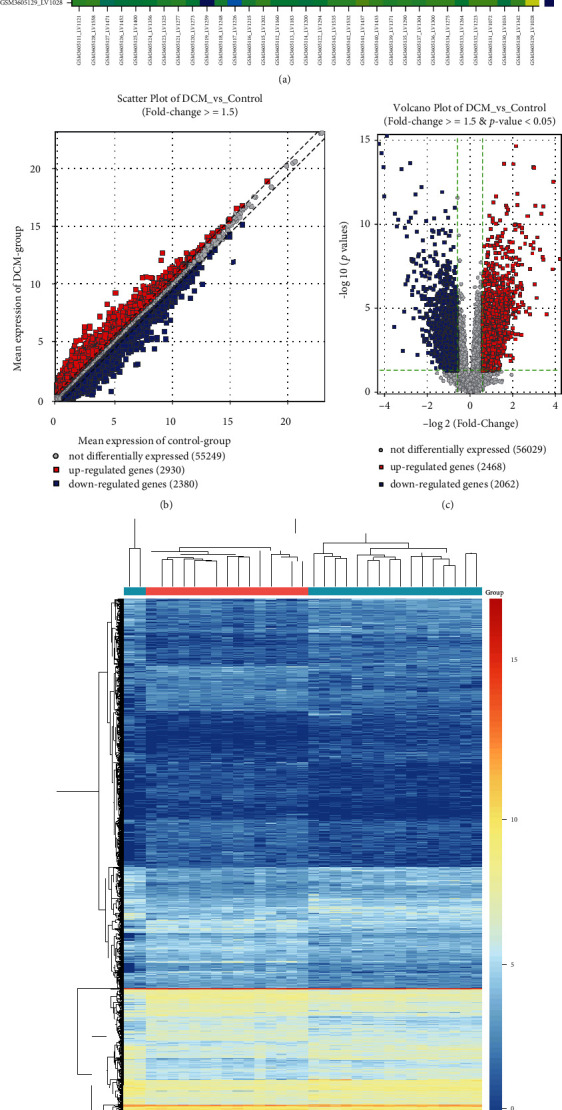
Figure 2.

Differentially expressed genes between DCM and control left ventricle specimens in the GSE126569 dataset. (a) Heat map for the correlation between DCM and control left ventricle samples. The closer to yellow, the higher the correlation coefficient. (b) Scatter plots showing up- and downregulated genes with ∣FC | ≥1.5 between DCM and control groups. (c) Volcano plots for DEGs with ∣FC | ≥1.5 and adjusted p value < 0.05 between DCM and control groups. (d) Heat map for DEGs between DCM and control groups. Red: upregulated genes; blue: downregulated genes; grey: no differentially expressed genes.
