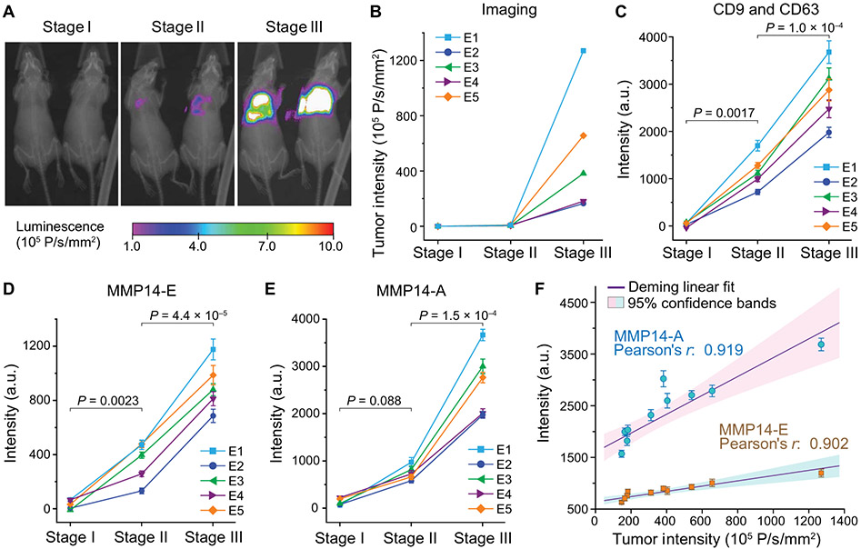
Fig. 4. Integrative sEV phenotyping for monitoring tumor development in mice using an experimental metastasis model of human breast cancer.
(A and B) An experimental metastasis model in athymic nude mice was established by injecting 106 2LMP-Luc cells into the tail veins of 4-week-old female nude mice. Progression of the lung tumors was monitored by bioluminescent imaging. (A) Representative images and (B) corresponding tumor intensity plots were acquired for each mouse at three stages: (i) before inoculation, (ii) initial detection of early metastasis, and (iii) nearly moribund with extensive lung metastases. (C to E) Multiplexed total sEV (CD9 and CD63), MMP14-E, and MMP14-A analyses of circulating sEVs in mouse plasma using EV-CLUE. Blood (~50 μl) was collected from tail vein at each stage. Plasma (6 μl) was diluted in PBS by five times and analyzed on chip using the anti-human mAbs validated for specific detection of human tumor xenograft-derived sEVs. Each sample was assayed in triplicate to determine the mean and SD (error bars). P values were determined by one-way repeated measures ANOVA with post hoc Tukey’s pairwise multiple comparisons test, P < 0.05. (F) Correlation of the sEV MMP14 expression and activity measured for 10 xenografted mice at stage III with the tumor sizes measured by bioluminescent imaging. All data were presented as means ± SD (n = 3). Deming linear fitting was performed at the 95% confidence level.

