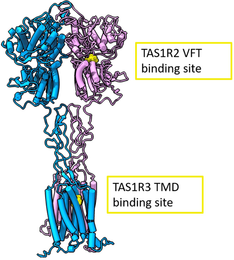
Fig. 1. Full human sweet taste TAS1R2/TAS1R3 receptor model with the TAS1R2 monomer colored in pink and the TAS1R3 monomer in cyan.

Binding sites are represented in yellow. The full receptor heterodimer was prepared with the I-Tasser web server51 based on multiple experimental structures (i.e., 6N51, 5X2M, and 5K5S). The binding site of TAS1R2 is based on coordinates of docked D-glucose to a Venus flytrap (VFT)59 (modeling based on template PDB ID: 5X2M, docking performed with Schrödinger Maestro 2019-1, Glide XP), and the TAS1R3-binding site is based on a lactisole molecule docked to the TAS1R3 TMD model (template PDB IDs: 4OR2 and 4OO9, Schrödinger Maestro 2018-2, Glide SP). The figure was made using ChimeraX (version 0.93)60.
