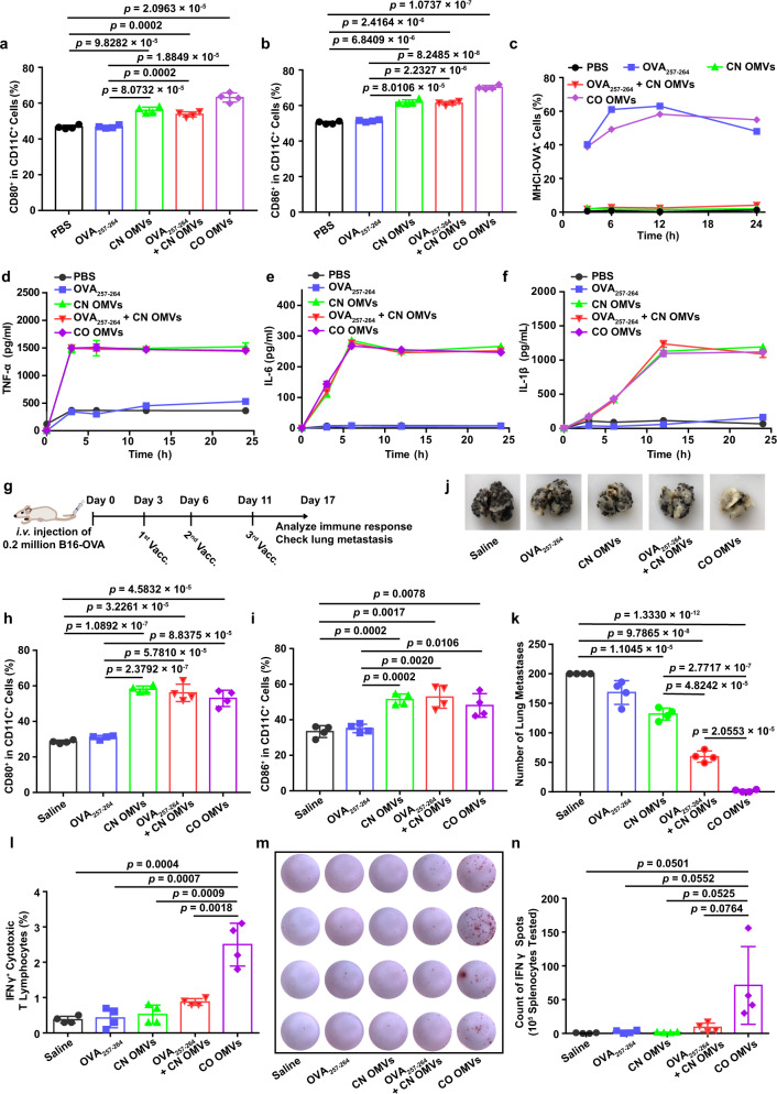
Fig. 2. Innate immune response and antigen-specific T-cell-mediated anti-tumor immunity induced by tumor antigen peptide-displayed OMVs.
a, b Maturation of BMDCs following treatment with OMVs preparations or controls. Flow cytometry was used to measure the percentage of CD80+ (a) or CD86+ (b) cells in CD11c+ BMDCs (n = 4). c The expression of the MHCI-OVA complex on the surface of BMDCs was measured by flow cytometry (n = 3). d–f TNF-α (d), IL-6 (e), and IL-1β (f) levels in the BMDC-conditioned medium after the indicated treatments (n = 3). g Schema showing the mouse B16-OVA melanoma model used to study the effects of OMVs vaccination (Vacc.) on lung metastasis. C57BL/6 mice were inoculated with B16-OVA melanoma cells (n = 4, 2 × 10⁵ cells/mouse, i.v.), then immunized with the following vaccines: saline, OVA257–264, CN OMVs (ClyA-none OMVs), OVA257–264 + CN OMVs or CO OMVs 3, 6, and 11 days later. Lung metastasis and immune responses were analyzed on day 17. h, i The maturation status of DCs in inguinal lymph nodes on days 17 post immunization. The percentage of CD80+ (h) or CD86+ (i) cells in CD11c+ cells was assessed by flow cytometry. j, k Lung metastasis was assessed on days 17 after tumor cell administration and following the indicated vaccine treatments. The lungs were photographed (j), and the tumor nodules in the lungs were counted (k). l Flow cytometry analysis of the percentage of IFNγ+ cytotoxic T lymphocytes (CD3+CD8+IFNγ+ T cells) in splenocytes re-stimulated with OVA257–264 antigen. m, n IFNγ secretion, as measured by the ELISPOT assay, from splenocytes which had been re-stimulated with OVA257-264 (m). Quantitative analysis of the ELISPOT assay for IFNγ secretion is shown in (n). g–n, n = 4. The data (a–f, h, i, k, l, n) are shown as mean ± SD. Statistical analysis was performed by a two-tailed unpaired t test. Source data are provided as a Source Data file.

