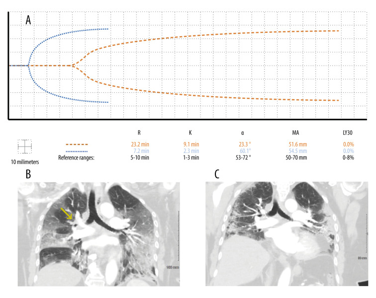
Figure 1.
Case 1: A 72-year-old woman with a past history of venous thromboembolism on chronic warfarin therapy presented with COVID-19 pneumonia. Three days after admission, she developed a pulmonary embolus despite an international normalized ratio of 6.4. (A) The patient’s initial thromboelastographic (TEG) tracing demonstrated hypocoagulopathy 1 day after developing the pulmonary embolus (dashed line). Two weeks later, her TEG tracing demonstrated hemostasis (dotted line). (B) Computed tomography angiogram of the chest in coronal view depicting branching filling defect along the distal right main pulmonary artery extending into the right upper lobe segmental branches (yellow arrow). (C) Right upper lobe pulmonary infarct was no longer present after repeat angiogram of the chest 2 weeks after its discovery.

