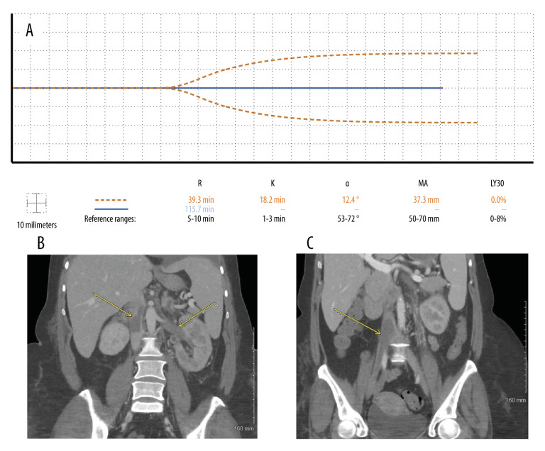
Figure 3.
Case 3: A 43-year-old woman with no past medical history was admitted with COVID-19 pneumonia, bilateral pulmonary emboli, and acute-onset idiopathic thrombocytopenic purpura. (A) Seven days after admission, the patient developed thrombi despite thromboelastography (TEG) depicting a nearly flat line tracing (dashed line). Two weeks later, the anticoagulant dose was increased after enlargement of venous thrombi, pushing her TEG tracing to a complete flat line (solid line). (B) Seven days after admission, computed tomography of the abdomen and pelvis in coronal view showed thrombosis within the entirety of the left renal vein and the inferior vena cava, both above and below the level of the renal veins. (C) Computed tomography 11 days later revealed enlargement of lower inferior vena cava and iliac thrombi.

