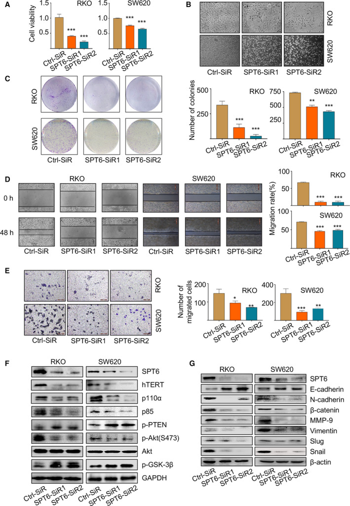
Fig. 2.

SPT6 promotes the proliferation and metastasis of colon cancer cells in vitro. (A) After knocking down SPT6, MTT assay was carried out to measure the cell viability in two colon cancer cell lines, RKO and SW620. ∗∗∗P < 0.001. (B) The impact of knocking down SPT6 on the morphology of colon cancer cells and the representative images are shown (n = 3). Scale bars represent 200 μm. (C) Colony forming assay was carried out in two colon cancer cell lines to evaluate the impact of knocking down SPT6 on the proliferation. Representative pictures are shown (n = 3). Scale bars represent 200 μm. Quantification of the colony forming assay was performed using image pro plus. Data are presented as mean ± SD (n = 3). ∗∗P < 0.01, ∗∗∗P < 0.001. (D) Wound‐healing assay was performed in two colon cancer cell lines to evaluate the impact of knocking down SPT6 on the migration. Representative pictures are shown (n = 3). Scale bars represent 200 μm. Quantification of the wound‐healing assay was performed using image pro plus. Data are presented as mean ± SD (n = 3). ∗∗∗P < 0.001. (E) Transwell assay in two colon cancer cells upon SPT6 knockdown. Representative pictures are shown (n = 3). Data are presented as mean ± SD (n = 3). ∗P < 0.05, ∗∗P < 0.01, ∗∗∗P < 0.001. (F) Western blot assay showed the impact on PI3K/Akt signaling pathway markers after knocking down SPT6 in two colon cancer cell lines. (G) Western blot assay showed the impact on EMT markers after knocking down SPT6 in two colon cancer cell lines.
