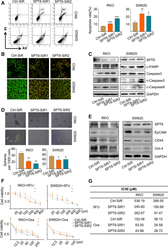
Fig. 3.

SPT6 knockdown induces apoptosis, stemness arrest and chemotherapeutic sensitivity improvement in colon cancer cells in vitro. (A) The apoptosis analysis of colon cancer cells upon SPT6 knockdown by flow cytometry based on FITC‐conjugated Annexin V/PI staining. The bar graph indicates the percentage of apoptotic cells. Data are presented as mean ± SD (n = 3). ∗P < 0.05, ∗∗P < 0.01, ∗∗∗P < 0.001. (B) The apoptosis analysis of colon cancer cells upon SPT6 knockdown by under a microscope following AO/EB staining. The cells with intact structures were stained green and were considered to be viable cells, whereas the cells with condensed yellow nuclei were identified as early apoptotic cells and those with condensed red‐orange chromatin were identified as late apoptotic cells. (C) Western blot assay showed the impact on apoptosis markers after knocking down SPT6 in two colon cancer cell lines. (D) The sphere‐forming assay was performed in colon cancer cells upon SPT6 knockdown as described in the methods section. Scale bars represent 500 μm. Quantification analysis of tumor spheres was performed using image pro plus. Data are presented as mean ± SD (n = 3). ∗P < 0.05, ∗∗P < 0.01. (E) Western blot assay showed the impact on stemness markers after knocking down SPT6 in two colon cancer cell lines. (F) Dose‐response curves of cell viability in RKO and SW620 cells transfected with control or SPT6 specific siRNAs and subsequently treated with varying concentrations of drugs for 48 h. (G) IC50 values of 5‐FU and Oxaliplatin in RKO and SW620 cells were treated as mentioned above.
