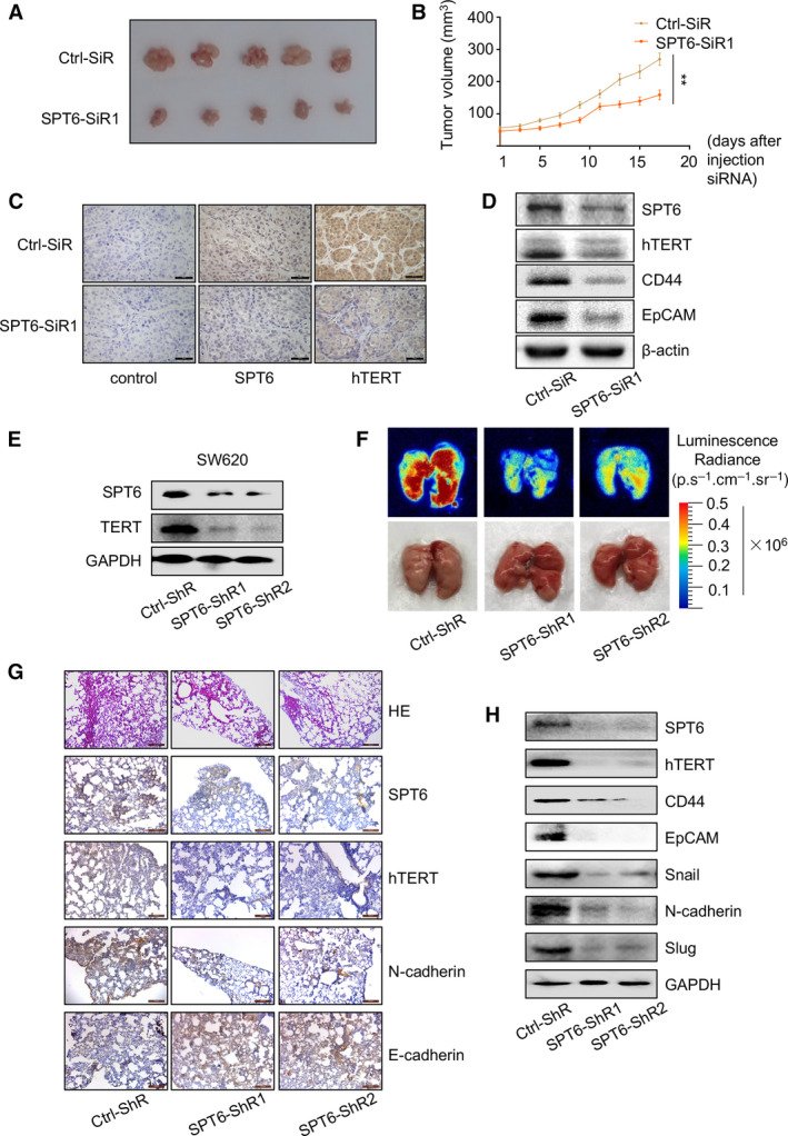
Fig. 4.

SPT6 knockdown inhibits tumor development and metastasis in mice. (A) The images of the tumors separated from the sacrificial mice on the 17th day after injection with control or SPT6 siRNA into the tumors. (B) The change of tumor volume upon knocking down SPT6 in the mice with xenograft of colon cancer cells. Data are presented as mean ± SD. ∗∗P < 0.01.(C) IHC staining of tumor sections collected from different treatment groups of mice at the end. Scale bars represent 100 μm. (D) Western blot assay of the expression of hTERT and stemness markers after knocking down SPT6 in tumors. (E) Western blot assay for the stable knockdown of SPT6 in SW620 cells. (F) The lung metastases of the mice assessed by in vivo fluorescence imaging and the corresponding apparent morphology imaging. (G) HE staining of the lung metastasis tissues in the mouse. Scale bars represent 200 μm and 100 μm). (H) Western blot assay of the invasion and stemness markers in lung metastasis tissues of mice.
